The femur is the bone in your thigh. It extends from your hip joint down to your knee joint.
It is one of the largest and strongest bones in your body, and because of this, it usually takes a very high energy force or injury to cause the femur to break (fracture).
If your bone is weak, for example in osteoporosis, your femur can fracture with a relatively minor fall.
Fractures of the femur are categorised by the exact location of the break (fracture):
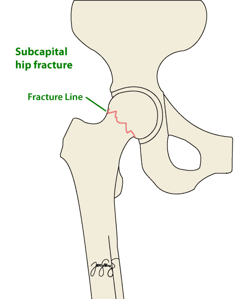
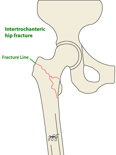
- Proximal femoral fractures. Proximal femur fractures, also called hip fractures, involve the upper-most portion of the thigh bone, just adjacent to the hip joint. These fractures are further subdivided into different types of hip fractures that have a bearing on how a surgeon may choose to fix the fracture.
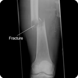
- Shaft fractures. A femoral shaft fracture is a severe injury that generally occurs in high-speed motor car accidents or falls from a height. The treatment is almost always with surgery. The most common procedure is to insert a metal rod down the centre of the thigh bone (an intramedullary rod).
- Distal femoral fractures. Also called a ‘supracondylar’ femur fracture, is an injury to the thigh bone just above the knee joint. These fractures can involve the cartilage surface of the knee joint, which put you at risk of developing knee arthritis later in life.
This section focuses on both shaft fractures and distal femoral fractures. For more information on proximal femoral fractures, see the section on hip fractures.


Symptoms
The symptoms of femoral fractures include the following:
- Pain and swelling: This will always be present in the instance of a femoral fracture. The location of pain will vary depending on the site of the break. For mid shaft fractures, the pain will usually be felt in the front of the thigh. For distal femoral fractures, the pain will be felt around the knee joint.
- Deformity: If the break goes right through the bone, there may be deformity of the thigh or knee. In a mid shaft fracture, the leg may appear shortened. For fractures close to the knee, the knee may appear to be at an abnormal angle.
- Numbness or weakness: the broken bone may put pressure on nerves travelling to the lower leg. This can lead to a feeling of numbness in the leg or pins and needles. It can also cause weakness of the lower leg muscles.
- Bruising or bleeding: The break in the bone can cause the blood supply to the thigh to be damaged. This can lead to widespread bruising over the thigh area. If the bone breaks through the skin (known as an open fracture) then there may be bleeding as a result of this.
In the case of a stress fracture, there will still be pain and swelling but not deformity, bruising or nerve damage. The pain and swelling will often come on gradually rather than immediately in the case of a fracture due to an accident.
Causes
There are three possible overall causes of femoral fractures:
High energy trauma

The femur is the biggest bone in the body and it is also one of the strongest bones. For this reason, a large amount of force is usually required to fracture the bone. An accident that causes this type of force is known as a high energy trauma. The type of accidents that can cause a femoral fracture in this way include the following:
- Motor vehicle trauma (eg, motorcycle accidents, motor vehicle accidents, plane crashes, pedestrian car accidents)
- Sports (eg, high-speed and contact sports with direct trauma, skiing, downhill mountain bike riding)
- Falls (eg, from height: mountain climbing, abseiling, workplace accidents)
- Gunshot wounds
Low energy trauma
If the femur is fractured due to a relatively small amount of force, such as a simple fall, we often suspect that there may be something that has caused the bone to lose some of its usual strength. These injuries that do not involve a lot of force are termed low energy trauma. The type of people that are at risk of fracturing their thigh bone after low energy trauma include the following:
- People who have decreased bone density due to osteoporosis. Elderly women are at greatest risk of this.
- People who have had cancer that has spread to the bones
- People who have been on long term corticosteroids. This has the effect of decreasing bone density leading to weaker bones.
Stress fractures
The third way to fracture the femur is through repetitive trauma. This creates a different type of fracture known as a stress fracture and usually comes on more gradually. It involves damage to the internal architecture of the bone but does not involve a clean break as in other femoral fractures. This occurs most commonly in athletes undergoing heavy training or military recruits. It is more common in women, particularly in women who are not menstruating. It is rare to have a stress fracture affecting the lower part of the femur. Most stress fractures of the femur affect the mid shaft area.
Risk Factors
The risk factors for sustaining a femoral fracture in a person with normal healthy bones involves activities that place them at higher risk of a high energy trauma and include the following:
- Motor sports (both car and motorcycle racing)
- Participation in high impact sports such as down hill skiing and down hill mountain biking
- Working or participating in sports at heights such as climbing or abseiling
The risk factors for sustaining a femoral fracture due to less severe accidents such as falls include the following:
- Osteoporosis
- Advanced age (particularly women)
- Long term corticosteroid use
- Previous cancer that is able to spread to bones
The risk factors for sustaining a femoral stress fracture include:
- Jogging frequently for long distances – particularly if training is not followed by appropriate rest times
- Women who are not menstruating (due to hormonal effects)
Investigations
Clinical assessment
The first step in diagnosing any problem is to obtain a thorough history of the problem at hand and perform an examination to determine what further tests may be required.
For femoral fractures, it is important that you give the doctor as much information about the accident that caused the bone to break. A simple fall at ground level as opposed to a fall whilst rock climbing will help determine what sort of fracture may be present. It is also important to let the doctor know of any medical conditions you have had in the past as well as the treatments you have received, even if they seem unrelated. For instance an asthmatic who has been on cortisone for many years may be at a higher risk of a fracture than someone who has never had to use cortisone.
An examination of the the whole lower leg (ankle, knee, hip and pelvis) will be carried out. As femoral fractures are often caused by accidents involving a large amount of force, other areas may be damaged as well as the thigh bone. The doctor will also assess whether the nerves and blood vessels of the lower limb are working properly or whether they have been affected by the broken bone. This can include the doctor testing skin sensation in the leg as well as muscle strength of the leg.
Imaging

Once an examination has been performed, it is usually necessary to take some radiographic images of the affected areas:
- X-rays: several X-rays may be taken of the femur and surrounding bones. This is an important first step in confirming that a fracture is present, but also the exact location and extent of the damage.
- CT scans: a CT scan may be necessary to give the doctors a clearer picture of the fracture. This is particularly important if surgery is required to fix the broken bone. The advantage of CT over X-ray is that it provides a 3D image of the leg and a more accurate picture of how far the fracture has spread, particularly if it affects the joint surfaces of the knee
- Bone scan: This test may be required if a stress fracture is suspected. Whilst X-rays can pick up some stress fractures, the bone scan is a more accurate tool to
Complications
The complications of a fractured femur vary depending on the location of the fracture and the severity of the break. The complications that can occur include:
- Infection: In the case of a fractured femur that results in bone breaking the skin, there is an increased risk of infection. This can be minimised with the appropriate use of antibiotics
- Bone healing Problems: If the bones are not well aligned or there is irritation to the bone due to infection, the healing process may be delayed and require further surgery
- Nerve damage: Nerve damage in femoral fractures is relatively rare but can lead to persistent numbness or weakness in the lower leg
- Compartment syndrome: This is a relatively rare complication of femoral fractures. It is the compression of nerves, blood vessels, and muscle inside a closed space (compartment) within the body. This occurs within the thigh when there is inflammation and bleeding due to the trauma associated with the fracture. If present, compartment syndrome requires an operation without delay.
- Surgical complications: These include failure of the hardware used to stabilise the bone or prominence of a piece of hardware leading to irritation and pain. Nerve damage is also a possible surgical complication.
Complications specific to the type of femoral fracture include:
- Distal femoral fracture: Due to the location of this type of fracture, the knee can be affected. There will often be stiffness of the knee which may resolve very slowly and may not fully resolve. Another way this type of fracture can affect the knee is by predisposing to osteoarthritis. This is most likely if the fracture line passes into the joint, disrupting the smooth layer of cartilage that lines the joint.
- Mid shaft fracture: This type of fracture can also affect the knee, but in a different way. Due to the movement of the femur when it breaks, there is often ligament damage to the knee which may require an operation in order to repair the damage. Mid shaft fractures in teenagers and children who are still growing may suffer leg length discrepancy where one leg is longer than the other. This can be due to the broken bone growing too much, or not enough, after the fracture.
Treatment
The treatment options for femoral fractures vary depending on the severity of the injury. Broadly speaking there are two main categories of treatment – conservative / non-surgical treatment or surgical treatment:
Non-surgical treatment
There are two non-surgical approaches. One is traction and the other is casting or bracing:
- Traction: This involves pulling on the part of the bone below the break to ensure that the two ends of the bone line up and will heal without deformity. It may be necessary to insert a pin in the lower leg bone in order to be able to apply this traction. Weights attached to pulleys are then attached to the pin in order to apply a constant level of controlled, appropriate tension to the affected bone.
- Casting and bracing: If the two ends of the broken bones are lined up well, it may be possible to simply apply a cast or a brace and wait for the bones to mend of their own accord. This approach can only be taken if there is good alignment of the bones following the break and there is not multiple pieces of broken bones.
NB: The draw back of both of these approaches is that the knee joint, and to a lesser extent the hip joint, are often immobilised for long periods. This can lead to stiffness of the joints and muscle wasting which can prolong a full recovery.
Surgical Treatment
The basis of surgical treatment is to fix the bones in place such that they line up as they normally should in order to allow proper healing. There are two main methods used in femoral fractures to attain this result:
- External fixation: This means that the bones are held in place using a metal frame that is outside the body with pins that then penetrate the bones. This approach is favoured where the fracture has lead to damage of the surrounding muscles and skin. It may also be favoured if the person who has sustained the fracture also has other medical conditions that would make a more invasive surgery difficult.
- Internal fixation: This approach means that the surgeon places supports around the bone on the inside of the leg. There are two main approaches used that come under the category of internal fixation:
- Intramedullary nailing. This involves a specifically designed rod to be placed through the centre of the bone shaft. The rod will cross the line of the fracture and keep the two ends of the bone together.
- Plates and screws: This involves the use of metal plates and screws to hold together the fragments of bone created by the fracture.
Joint replacement
In the case of a severe lower femoral fracture, the bone may not be able to be effectively stabilized using the techniques previously mentioned. Instead, it may be necessary to replace the section of damaged bone with an implant leading to a partial joint replacement.
Seeking Advice
A fracture of the femur is a serious injury and requires urgent medical attention. If a fracture of this kind is suspected, it is important to not try to stand on the affected leg. Ask someone to call an ambulance so that you can be safely transported to the hospital where you can seek appropriate medical help.
If you suspect a stress fracture however, this can be initially managed by your GP who will be able to decide if you need a specialist consultation.
Prevention
High energy accidents that result in thighbone fractures are generally not foreseeable, so can’t be prevented!
For more information on prevention of fractures resulting from osteoporosis, click here.
