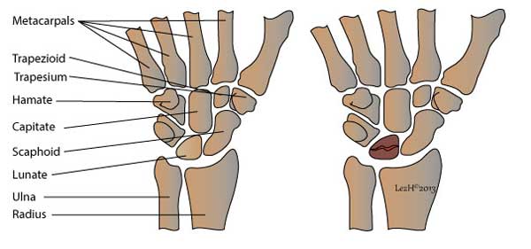
Bone is a living tissue and requires continuous blood supply for nourishment. Kienbock’s disease is a condition where one of the eight bones (lunate) in the wrist, loses its blood supply and thus causes death of the bone – this is known as osteonecrosis.
There are 2 main rows of bones that make up the wrist bone. The front row (proximal) includes scaphoid, lunate, triquetrum and pisiform. The back (distal) row consists of trapezium, trapezoid, capitates and hamate. The front row of bones articulates with the 2 forearm bones – the radius and ulna, to form the portion for wrist motion.
Damage to the lunate bone can lead to persistent pain, swelling and stiffness. Kienbock’s disease tends to occur more in men between the ages of 20 – 40 and rarely affects both wrists. Patients usually have a history of trauma to the wrist.

Symptoms
Patients with Kienbock’s Disease will develop the following symptoms:
- Pain and swelling around the wrist
- Pain or discomfort directly on the bone itself
- Stiffness of the wrist
- Reduced strength in the wrist.
- Difficulty in turning the hand upward usually due to pain

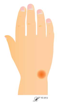
Diagram showing the location of pain of Kienbock’s disease
Causes
The exact cause of Kienbock’s Disease is not known. It might involve several factors such as the blood supply to the bone (artery), blood drainage (veins) and skeletal variations (anatomy). Skeletal variations include the shape / size of the lunate, and the shorter end of the ulnar (forearm bone) may predispose patients to developing this condition. Traumatic insult and repetitive mechanical loading may possibly be a factor in some cases. Occasionally, Kienbock’s disease is associated with certain conditions such as septic emboli, sickle cell disease, gout and cerebral palsy.
Risk Factors
Risk factors for the development of Kienbock’s Disease include:
- Occupations that require the use of pneumatic tools such as rivet guns and hammers – increased impact loading on the wrist
- Abnormal wrist posture – this will compromise blood supply to the lunate.
- Repetitive trauma to the wrist.
Investigations
Your doctor may perform the following tests, which include:
- Physical Examination: Your doctor will apply pressure on the lunate to elicit any pain or discomfort. He / She will also assess the range of movements of the wrist.
- X-rays: May be useful to determine how far the disease has advanced. In early stages, the x-rays may appear normal.
- Others: Your doctor may recommend using MRI, CT scan and bone scan. MRI is reliable in accessing the blood supply of the lunate.
Kienbock’s Disease progresses through four stages. In early stage, it is difficult to diagnose Kienbock’s Disease because the symptoms are very similar to a sprained wrist.
Stage 1: Symptoms are similar to those in wrist sprain. X-ray may be normal or show features of fracture. MRI may be useful in diagnosis in this early stage.
Stage 2: Pain and swelling of the wrist is common in this stage. Lunate bone begins to harden (sclerosis). Dense white areas on x-rays indicate that bone is dying. CT scan or MRI is able to assess the bone.
Stage 3: The dead bone begins to collapse, and surrounding bones may start shifting position. During this stage, there is increasing pain, motion restriction of the wrist and weakness in gripping.Stage 4: The structures surrounding the lunate may be affected. This will result in arthritis of the wrist.
Complications
Failure to treat the condition may result in degenerative changes around the joints, increasing the risk of developing wrist arthritis. Severe stiffness and motion loss of the wrist will interfere with daily activities.
Chronic wrist pain may progress to regional pain syndrome.Other complications include: muscle weakness and instability of the wrist.
Treatment
Treatment can be non – surgical or surgical. The goal is to restore blood flow to the lunate and relieve any pressure on the lunate.
Non – Surgical: The wrist may be splinted for about two to three weeks. Your doctor may also prescribe some anti – inflammatory medication such as, ibuprofen and aspirin to reduce swelling and pain.
Surgical: Your doctor may recommend surgical treatment if the symptoms do not resolve. Surgical methods include:
- Re-vascularization: this procedure involves taking a bone graft from the inner bone of the lower arm to restore blood supply to the lunate.
- If the forearm bones are uneven, a levelling procedure can be done. Bone can be made longer or shorter by using bone grafts. This levelling process prevents the bear down on the lunate and slows the progression of this condition.
- Metal devices can be used to preserve the spaces between bone and to relieve pressure on the lunate.
- In severe cases where the lunate is collapsed, this can be removed. Two adjacent bones are also removed to maintain partial wrist motion and relieve pain – proximal row carpectomy.
- Another procedure is done where several bones are fused together – arthrodesis. This is usually done in wrist arthritis to reduce the pain and help maintain function. However, the range of motion will be limited.
The choice of procedure depends on several factors: disease progression, staging, personal goals and activity level.
Seeking Advice
Your Family Doctor (GP)
Your Family Doctor will be able to diagnose and help treat your problem. He or she will be able to
- tell you about your problem
- advise you of the best treatment methods
- prescribe you medications
- and if necessary, refer you to Specialists (Consultants) for further treatment
Prevention
There isn’t a clear and define way to prevent Kienbock’s Disease. It may help if you:
- Avoid repetitive trauma to the wrist – wear protective equipments during sport.
- Treat the underlying cause – septic emboli, sickle cell disease.
F.A.Q. | Frequently Asked Questions
What is the prognosis?
The treatment results vary considerably, depending on the severity of involvement and whether or not the disease progresses. The response to treatment can take several months. Occasionally, multiple treatments or surgeries might be necessary.
What happens after surgery?
The wrist is splinted for about 12 weeks after surgery. Your surgeon will x-ray the wrist several times to make sure the bone is healing and alignment is correct. Once your wrist has begun to heal, your doctor will recommend you to begin a rehabilitation program.
Rehabilitation
You may need both physical and occupational therapy sessions. These sessions include exercise to help strengthen muscle around the wrist joint, improve fine motor and dexterity of the hand. Advice will also be given to prevent extra strain on your wrist joint while doing your daily activities.
