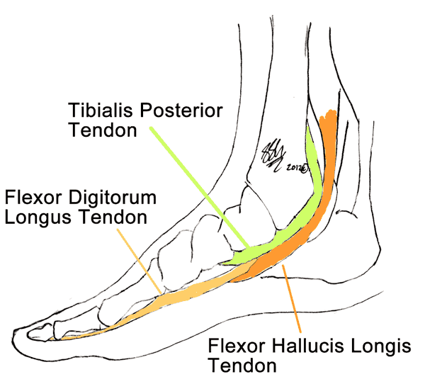
Tibialis posterior dysfunction is a broad term describing injury or disease in the tendon of one of the muscles in the calf. This disorder is also known as posterior tibialis tendon dysfunction, or PTTD, and posterior tibialis tendonitis, tendonosis or tendinopathy, and posterior tibialis insufficiency. All of these terms describe different types or degrees of injury to the tendon resulting.
The posterior tibialis muscle is found deep on the back of your calf. It begins near the knee, joins its tendon just above the ankle and runs down to attach to a bone (navicular) on the inner surface of your foot. This allows it to support the instep arch, a key component of normal, painless walking. This muscle is also partly responsible for pointing your foot down (‘plantar flexion’) and in (‘pronation’).

Tibialis Posterior dysfunction refers to injury or disease affecting the tendon, the strong band of tissue that connects muscle to bone. Injury to the tendon occurs when the tendon is overstretched or strained. Lots of tiny tears in the tendon results in inflammation – a process called tendinitis.
There are a number of different symptoms associated with PTTD, including pain along the inner side of the lower leg, ankle or foot. PTTD is also associated with the development of flatfoot deformity.
Symptoms
Early symptoms of PTTD include redness, swelling and pain along the course of the tendon (the inner surface of the foot and ankle). This may result in pain walking or running, and pain lifting the foot or weight-bearing on the balls of the feet. Your doctor may notice that your feet flatten (over pronate) during walking, though this is probably not something you will be able to see yourself.
Later symptoms reflect the impact of poorly balanced muscle acting around the ankle and foot. You may notice your foot gradually deforming or twisting and decreased muscle strength.
Surgeons and doctors use a series of stages to assess the severity of PTTD. There are four stages:
- Stage 1– the inflamed tendon is painful and swollen along the inner surface of the ankle.
- Stage 2– Pain is more severe and the foot gradually flattens as the arch begins to collapse.
- At this point, you may notice you struggle to stand on you toes and your heel seems to roll outwards as you walk.
- Stage 3– By this point, you may be experiencing pain all over your foot. Your arch will now be fixed flat, and your heel rolls out more obviously as you walk.
- Stage 4– Once you have reached stage 4, your PTTD is affecting your ankle, causing it to become painful, swollen and stiff from the strain. You will also have more exaggerated deformity in the foot itself.
Most sufferers of PTTD do not progress all the way through to stage four.
Causes
Posterior tibial tendonitis occurs when the tendon has been strained by overuse. When the tendon is injured the arch of the foot flattens, resulting in extra strain pulling through the tendon. This sets up a vicious cycle, with further injuries increasing the likelihood of longterm problems.
Alternatively, some people are born with flattened arches, or develop flatfeet from other causes. This again causes excessive strain through the tendon during walking and running, and makes the tendon vulnerable to injury.
Risk Factors
People at increased risk for developing tibialis posterior dysfunction include:
- While you can develop PTTD at any age, middle-aged women are the most likely to develop symptoms.
- Flat feet- Many people who develop this condition, already have flat feet. Overuse or constantly high demands on the tibialis posterior tendon can cause it to become inflamed and irritated.
- Other factors- obesity, other medical conditions including hypertension and diabetes.
Investigations
You will have an examination of your foot and ankle by a doctor or specialist. X-rays are used for diagnosis and to assess the arthritis and alignment of the joints in the foot and ankle. You may also need an X-Ray of your other foot as a comparison.
Sometimes ultrasound, CT or MRI scans are used to confirm the presence of inflammation in the tendon.
If your doctor is concerned about rarer causes of tibialis dysfunction, he or she may order some blood tests.
Complications
There are a number of complications that may develop. This includes:
- Deformity. Over many years of PTTD bony and soft tissue structures supporting the foot start to warp and degenerate. This leads to two common types of deformity, equinus and varus. In equinus deformity the foot points down, like a ballerina. In varus deformity the foot points inward. Either or both of these may occur in PTTD and require may require correction to facilitate walking or reduce pain. Many of the special supporting footwear used in PTTD treatment aims to prevent the development of deformity.
- Rupture. An inflamed, irritated tendon is at risk of tearing completely, or rupturing. This is a very painful experience, accompanied by severe redness and swelling. The tendon can be repaired with surgery.
Other complications include arthritis in other joints involved in walking (like hips, knees and lower back) and ulcers or callouses developing where the deformed foot rubs
Treatment
There are non-surgical and surgical options to treat Tibialis Posterior Tendon Dysfunction. The chosen management will depend on the severity of your condition and the likely progression of symptoms in the future. This will be assessed and discussed with you by your doctor or surgeon.
There are many treatments for tibialis posterior dysfunction that do not involve surgery. They are often very successful, incorporating medications, exercises and physical support of the foot. The aim of these approaches is to:
- Rest the tendon, relieving the strain of weight-bearing.
- Retrain the muscles and ligaments around the ankle and foot to prevent re-injury or worsening of the dysfunction.
Non-surgical treatments recommended for most sufferers of PTTD includes:
- Rest. When a tendon is inflamed, it needs rest in order to recover. This can mean not walking, walking with elbow crutches or walking with a special boot that minimises the weight bearing through the tendon itself. The absence of strain allows the inflammation to die down so that pain and swelling decrease, and facilitates healing. Your Consultant will tell you if this is appropriate for you and how to rest.
- Ice. Applying ice packs to your foot for 20 to 30 minutes every 3 to 4 hours for the first 2 to 3 days or until the pain goes away. Thereafter, ice your foot at least once a day until the other symptoms are gone. Ice massage – Freeze water in a cup and then peel back the top of the cup. Massage the ice into the painful tendon for 5 to 10 minutes.
- Elevation. Keeping your lower leg and foot above the level of your chest helps drain blood and fluid out of the leg, reducing swelling. Sleeping with your foot on a number of pillows is a great way to keep your leg elevated and reduce swelling.
- Medication. Painkillers and anti-inflammatory medications will help with the pain and inflammation. These will therefore help to reduce the discomfort.
Footwear and physical therapy are also very successful ways of treating posterior tibialis tendinitis.
- Foot wear. Wearing good shoes can help minimise discomfort from Tibialis Posterior Tendon Dysfunction. This includes shoes that are stiff and supportive. You will know if your shoe is sufficiently stiff if when you hold it in your hands, it is difficult to bend in half. Lace up boots that limit the movement of the ankle as well are very helpful. Weight loss The tendon’s inflammation will be greatly reduced if your body weight is within a normal range.
- Orthotics. There are several orthotics options for the treatment of Tibialis Posterior Tendon Dysfunction. These can be either insoles or a more sturdy ankle support. Both of these types of orthotics will fit in your shoes to help your foot and ankle function better.
- Taping. An orthoptist or podiatrist may recommend placing strong tapes across the sole of the foot to support the arch.
- Plaster Cast. Occasionally, your doctor may put a cast on your ankle to prevent weight-bearing and rest the tendon. This will usually be for only a short time.
- Physiotherapy treatment to reduce inflammation of the tendon, improve muscles’ flexibility and strength can improve the pain. Exercises prescribed by physiotherapists can help speed recovery and return to normal activities, including sports.
If non-surgical treatment does not improve pain and deformity then surgery may be considered.
What are the surgical options for Tibialis Posterior Tendon Dysfunction?
There are several types of surgery for the treatment of Tibialis Posterior Tendon Dysfunction. These options include:
- Tendon Debridement. This involves removing inflamed and irreversibly damaged parts of the tendon. It can be very effective in pain relief for mild tendon inflammation where the bones and arch of the foot are still normal.
- Achilles tendon lengthening. A tight or short Achilles tendon contributes to the symptoms of PTTD, and a simple correction of this may be enough to relieve pain.
- Tendon grafts. Your surgeon may be able to harvest tendon from other parts of your foot, usually the flexor digitorum longus tendon. This can then be used to reinforce the tibialis tendon. This operation can relieve pain as well as improve walking.
- Osteotomy. Osteotomy is a term used to describe reshaping of the bones.
- Corrective fusion of hindfoot. This is a drastic form of surgery, usually only recommended as a last resort. More information about this form of surgery can be found in Arthrodesis of the Foot.
For moderately severe PTTD, surgeons may use a combination of Achilles lengthening, grafts and osteotomy to restore function to the foot.
Recovery from tendon debridement is fairly rapid. In contrast, rehabilitation from the more involved repair surgery may be long, involving an intense physiotherapy regime as the tendons and bones heal.
Seeking Advice
If you have persistent pain while walking in spite of rest and over the counter pain relief, you should see your doctor.
Your Family Doctor (GP)
Your Family Doctor will be able to diagnose and help treat your problem. He or she will be able to
- tell you about your problem
- advise you of the best treatment methods
- prescribe you medications
- and if necessary, refer you to Specialists (Consultants) for further treatment
Prevention
Maintaining a healthy weight and wearing comfortable, well-fitting shoes can help protect your feet.
Arch supports are a great way of limiting the progression of PTTD.
F.A.Q. | Frequently Asked Questions
When can I return to my normal activities?
Everyone recovers from an injury at a different rate. Return to your activities depends on how soon your injured tendon recovers, not by how many days or weeks it has been since your injury has occurred. In general, the longer you have symptoms before you start treatment, the longer it will take to get better. The goal of rehabilitation is to return you to your normal activities as soon as is safely possible. If you return too soon you may worsen your injury
You may safely return to your activities when, starting from the top of the list and progressing to the end, each of the following is true:
- You have full range of motion in the injured leg and foot compared to the uninjured leg and foot.
- You have full strength of the injured leg and foot compared to the uninjured leg and foot.
- You can walk straight ahead without pain or limping.
References
Beals, T. C., et al., ‘Posterior Tibial Tendon Insufficiency: Diagnosis and Treatment’, Journal of the American Academy of Orthopaedic Surgeons, Vol. 7, No. 2, March-April 1999, pp. 112-118.
Haendlmayer, K. T., Harris, N. J., ‘Flatfoot deformity: an overview’, Orthopaedics and Trauma, Vol 23., No 6., pp 395-403.
Weinraub, G. M., Saraiya, M. J., ‘Adult flatfoot/posterior tibial tendon dysfunction: classification and treatment’, Clinics in Podiatric Medicine and Surgery, Vol. 19, 2002, pp 345-370.
