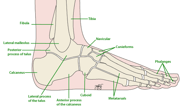
The talus is the in your foot that forms your ankle joint with the leg bones. It is the bone in the foot directly supporting the leg.
Usually a great deal of force is required to cause fractures or a dislocation of the talus. This can include falls and car accidents, as well as sports like snow-boarding. However, sometimes talus fractures occur with only small injuries when the bones are weak, as in osteoporosis.
Fractures of the talus are often difficult to manage. Most of the bone surfaces are covered by a protective smooth substance called cartilage. Cartilage does not allow blood vessels to enter the bone, and this means that the talus has a vulnerable blood supply. For this reason, talar fractures are frequently associated with death of the bone, or ‘avascular necrosis‘.
Talus fractures will often require surgery and long rehabilitation. Severe fracture-dislocations of the talus sometimes require removal of the bone fragments and ankle fusion (arthrodesis) to achieve recovery.

Symptoms
A fractured talus may cause the following symptoms:
- Pain & difficulty weight-bearing
- Swelling & tenderness when the foot is touched
- Bruising
- Deformity – the foot might appear as though it is shaped differently to the uninjured foot due to the fracture
Causes
As with many types of fractures, the causes and risk factors can be divided into two types:
- Large forces on strong bones
- Talar fractures are common among sports people who sustain high impact falls and trauma. The sport commonly associated with talar fractures is snow-boarding.
- Other common causes are car accidents and falls onto the feet.
- Small forces on weak bones
- Osteoporosis and advanced age
- Medications and other drugs; most commonly, people who need to take high doses of steroids for long periods of time are at risk of developing weak bones
Risk Factors
Risk factors for talus fractures include:
- Playing high impact, high speed sports, like snow-boarding
- Weak bones – usually due to osteoporosis
Investigations
Talus fractures will often require both X-Rays and CT scans to confirm the fracture and assess the degree of injury to the surrounding tissues.
Subtle fractures in the talus can be very difficult to see immediately after the fracture has occurred. In later weeks, the healing reaction of the bone can be more easily seen on X-Ray. For this reason, you may require x-rays a few weeks apart to diagnose the fracture.
Complications
Complications vary depending on the type and site of the fracture. Some relatively common complications at the time of injury include:
- Infection. This is especially likely to occur if the skin around the ankle has been broken during the accident. Your surgeon will give you antibiotics to help prevent infection.
- Stiffness in the foot and ankle. The talus is a key bone in the ankle and foot, and while it is healing you may have to keep it still for weeks at a time. This unfortunately makes stiffness in the foot and ankle increasingly likely.
- Injury to other components of the foot. The accident that causes the talus fracture may also cause sprains and tears in the tendons, muscles and other tissues around the talus.
There are a number of possible problems or complications related to any surgery – more information on these can be found here.
Some long-term complications include:
- Long-term pain, deformity and/or stiffness in the foot. The talus plays a key role in both the ankle and foot and consequently, damage to this bone is likely to affect both these areas. It is not always possible to predict who will develop these symptoms or how severe they will be. When pain is significantly affecting daily activities, a ‘salvage’ surgery can be used to relieve pain. This is most often arthrodesis, or fusion, of the joints.
Some rarer complications include:
- Avascular Necrosis (AVN) – this literally means death of the bone due to inadequate blood supply (a similar term is Osteonecrosis). It is a significant risk in talus fractures because the blood supply to the talus is relatively vulnerable. AVN is a serious complication because in addition to pain and instability, the dead bone is likely to become infected. For this reason, the dead bone needs to be removed. Some people are at greater risk of developing osteonecrosis in the talus, including those with diabetes, poor blood vessels to their foot, smokers and the elderly.
- Nonunion and malunion. These two occur when the bone fails to heal or heals in an inappropriate position. It sometimes occurs when weight is put on the healing bone too early, but can occur for no apparent reason.
Treatment
In mild talus fractures where the bones remain in place, the foot can be immobilised in a cast without surgery. However, surgery is required in most forms of talus fracture. Following surgery, most people will need to avoid putting any weight on their foot for long periods of time (see Non-Weight-Bearing).
Outcomes following talus fracture will depend greatly on whether the bone survives or becomes infected and needs to be removed (see Complications of Surgery). Even when the rehabilitation period is complication-free, rates of osteoarthritis following the injury are high.
What may be involved in the operation?
The type of fracture and severity of injury to the surrounding tendons will influence the type of surgery chosen. Your surgeon will be able to discuss this with you in more detail, however some aspects that may come up include:
- ‘Open reduction and internal fixation’. This term is commonly used by orthopaedic surgeons, and just means that the skin will have to be cut (‘open’) to reach the injured bone, and then screws need to be drilled across the fracture to keep in the injured bone in place (‘fixation’). While there a number of different techniques used to fix talus fractures, a common approach is to fuse the bones together (see fusion operations, ankle fusion and foot fusion).
- Bone grafts – if the talus is so damaged that fragments of bone need to be removed, your surgeon may ‘harvest’ extra bone from a thin bone in you leg called the fibula.
What happens in the first week after surgery?
After your operation, you will wake up with a large cast or bandage on your foot. For the first few days, you will need to keep your foot elevated and leave the cast in place to let the bone heal. After a few days, this cast is removed and a new cast applied. With this cast you will be asked to keep your foot elevated as much as possible (to prevent swelling and avascular necrosis), but also to perform exercises to keep the joints and muscles in you leg supple.
During this time you may be kept in hospital depending on
- the severity and type of your injury,
- your other medical conditions (for example, if you have heart disease)
- and on your level of support at home.
If you remain in hospital you may be required to wear special thick white stockings and take drugs to prevent clots forming in your leg (see DVT). Once your doctors think you’re safe to return home, you may be reviewed by occupational therapists.
If you notice any of the following symptoms while in hospital inform your doctor immediately:
- Severe pain, or pain that gets worse in the days following the operation
- Tingling and numbness in your toes
- Toes that turn blue
Physiotherapy is vital in rehabilitation after talus fractures, so you will be seen by a physiotherapist either during your stay in hospital or very soon after you are discharged. They will advise you on exercises that will facilitate healing and maintain the other muscles and joints in your leg. During your rehabilitation, you will have a series of appointments with your physiotherapist who will:
- Review your joint and muscle function
- Assess your walking pattern
- Recommend exercises to promote the healing of your bones and muscles, as well as maintaining strength and flexibility in the other joints in your leg
For more information, see the ‘Exercises’ section in General Aspects of Physio Rehabilitation.
- NOTE – do not, unless otherwise advised, perform passive range of motion exercises. These involve moving your foot with your hands, or asking someone else to stretch your foot for you.
And then?
At around two weeks you will be reviewed by your surgeon who will:
- Remove your cast and check your ankle and foot for signs of skin damage and nerve and blood vessel injury
- Remove any stitches
- Check the joints in your foot are moving and not painful
- If you have not already had a review X-Ray, you will be asked to get one at this point. The review X-Ray lets your surgeon check that your bone is in the correct position and is healing well. (Keep in mind that this X-Ray does not exclude the possibility of avascular necrosis developing later on.)
At this point, you should still be keeping your leg elevated as much as possible. If your foot becomes very swollen the skin and blood supply can be compromised.
Your surgeon will now suggest that you start putting some weight through your injured foot by placing your toe on the ground. This is called ‘partial weight-bearing’ and it is important that you:
- Only weight bear with assistance – for example, while leaning on a table or chair that can carry your weight. Your physiotherapist may recommend using crutches or other gait aids in these exercises.
- Do not put more weight on your leg than you can handle. If weight-bearing causes you significant pain do not force yourself.
Weight-bearing at this point helps to maintain the muscles in your leg and keeps your joints flexible.
After four weeks
You will again be reviewed by your surgeon who will check or change your cast or walking-boot, assess your foot and ankle and get review X-Rays. At this point, your surgeon may be able to tell you if your bone graft is healing into the talus and if the blood supply is good.
Your physiotherapist will increased the types of exercises you can do to include strengthening exercises as well as extending your range of motion and flexibility exercises.
If healing is progressing well, you can begin more weight-bearing. This usually involves putting some of your weight on your foot while walking with crutches. Do not, at any time, put your entire weight through your foot.
After six weeks, review X-Rays and an appointment with your surgeon will help to determine if your cast can be removed permanently and if your bones are healing well.
After two to three months
Depending on how well your bones are healing on X-Ray, you may be able to start full weight bearing. Do not force yourself to take on more than is comfortable. At this point, you may still require walking aids to help you get around.
