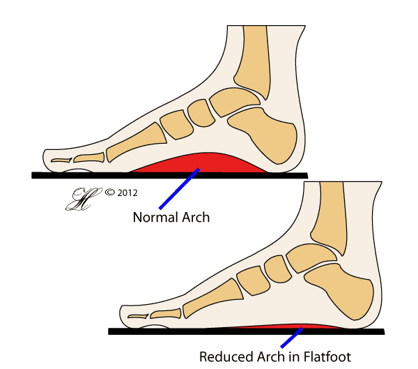
The normal foot can come in many shapes and sizes. Some people have a high arch, whilst others can have a very low arch on the inside aspect of the foot. All these variations are normal, as long as your joints are painless and you can move your foot normally.
The term ‘pes planus’ refers to a flattening of the inner arch of the foot (or medial longitudinal arch). The loss of this arch results in the two secondary components that make up pes planus – outward twisting of the heel (‘hindfoot valgus’) and outward deformity of the end of the foot in comparison to the ankle (‘midfoot abduction’).
If you have a flatfoot that is not causing you pain or any other problems, then the good news is that you have nothing to worry about. No treatment at all is needed. However, some people with flatfeet have an underlying problem that needs treatment. These problems may include:
- Congenital deformities. Some people are born with conditions which may lead to flat foot.
- Tibialis Posterior Dysfunction. This condition commonly affects women over 40 years old. The tibialis posterior muscle and tendon helps maintain your foot arch and helps you walk efficiently. When it tears, a flatfoot develops which may lead to osteoarthritis of your foot if not treated properly.
- Arthritis of the Joints in the Foot. Rheumatoid arthritis or other types of arthritis can affect your midfoot, leading to painful flatfoot.
- Neuromuscular Disorders. Conditions such as Polio, Charcot-Marie-Tooth Disease or Diabetes can cause Flatfoot.

This picture is a diagrammatic representation of flatfoot disorder. In reality the flattened effect occurs when ligaments that make up the arch slacken.
Symptoms
When flatfoot deformity becomes problematic, symptoms may include:
- Increasing pain in the foot – the pain is usually located on the inner surface (posteromedial hindfoot pain) and is worse when walking or standing still for long periods of time.
- Occasionally, there is also pain on the outer surface of the ankle near the lateral malleoli. This is due to the flattened arch causing bones to rub against each other abnormally (fibula impinging on the calcaneus).
- Swelling
- Abnormal gait – the flattened arch can make running difficult, even impossible. Walking may also be affected, especially when there is significant pain.
- A painful, abnormal gait will start to impact on the other joints involved in walking, namely the knees, hips and lower back. For this reason, secondary pain and arthritis may develop in these joints. Sometimes, lower back pain may be the only symptom of flatfoot deformity.
- Stiffness in other joints in the foot. With severe flatfoot deformity, the subtalar and transverse tarsal joints can become less flexible and even fixed in position.
- Associated deformities in the foot. The joints in the foot and ankle all work together, so when one is affected it is unsurprising that the others are also deformed. Any of the joints in the foot may be affected, however a common deformity is equinus deformity of the ankle (when the foot tends to point down).
- Associated conditions in the foot – plantar fasciitis, tarsal tunnel syndrome.
Causes
Frequently there is no recognised cause for flatfoot deformity. However some causes include:
- Other diseases:
- Posterior tibialis dysfunction.
- Rheumatoid arthritis and other forms of inflammatory arthritis (including Reiter’s syndrome, psoriatic arthritis, ankylosing spondylitis)
- Trauma. Lisfranc fracture-dislocations are associated with flatfoot deformity, however these are rare. Other forms of trauma affecting the talonavicular joint (an important joint in the centre of the foot) are also potential causes of flatfoot.
- Neurological causes that lead to a ‘Charcot foot’. The term Charcot foot refers to deformity that occurs when nerve disease leads to abnormal bones and unnoticed injury. Diseases that can produce a Charcot foot include diabetes and spinal cord injuries.

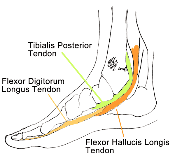
The arch of the foot is supported in part by the actions of these three muscles (found in the leg and stretching down around the ankle). In particular, weakness in tibialis posterior results in flatfoot.
Risk Factors
- Family history of flatfoot
- Childhood flatfoot
- Other forms of arthritis, including rheumatoid and the seronegative arthritides (psoriatic arthritis etc)
- Obesity
- Diabetes and Charcot foot
- An overly flexible foot
Investigations
Your GP or primary healthcare provider will often be able to diagnose flatfoot just by looking and feeling your foot, however the multiple other deformities and conditions associated with flatfoot may demand further tests. These may include:
- X-Rays – it is often helpful to look at weight-bearing X-Rays of the affected foot, and sometimes even to compare your feet to each other. Weight-bearing X-Rays show the alignment of the bones and can therefore be used to determine whether the foot is deformed.
- CT and MRI – these tests are occasionally used to give greater detail about the bones and the soft tissues in the foot and ankle. They are usually not necessary to diagnose flatfoot but may be useful in assessing severity or for finding the underlying cause, for example tarsal-calcaneal coalition.
- Blood tests – your doctor may order these to check if you have other conditions, for example rheumatoid arthritis, that cause flatfoot.
Sometimes it is helpful to look at other joints, especially the knee, hip and lower back, to assess whether the flatfoot deformity is affecting other parts of the body.
Complications
The main complication of concern in flatfoot deformity is posterior tibialis dysfunction.
Other complications include:
- Arthritis in the feet or other joints (knee, hip and lower back)
- fixed deformities in other joints.
- Infection / ulcer developing where the shoes rub against the abnormal foot
Treatment
The first step in treating flatfoot is:
- Relieving symptoms
- Pain and swelling can be relieved with over-the-counter pain medications (eg nurofen).
- Weight loss can help relieve symptoms by reducing the amount of force through the foot.
- Comfortable and supportive footwear
- Preventing worsening of the deformity and the development of associated conditions (especially posterior tibial tendon dysfunction).
- Orthotics are often extremely helpful in this regard. Both soft and rigid orthoses may be recommended.
- Ankle braces can help take some of the load off the foot.
At this stage, your doctor may refer you on to see a podiatrist and/or an orthotist.
When the above ‘conservative’ measures fail to relieve symptoms, surgery may be necessary. There are many surgical options available that fall into the following broad categories:
- Soft tissue surgery.
- As irritation of the posterior tibial tendon is a part of flatfoot deformity, surgery on this tendon can help relieve symptoms and correct the deformity. Grafts taken from other tendons may be used to lengthen the tendon.
- Ligament reconstruction. There are a few different ligaments that can be repaired or reconstructed including the medial deltoid ligament and the ‘spring’ ligament. Repair of these structures helps to recreate stability around the joints.
- Achilles and calf muscle (gastrocnemius) surgery.
- Osteotomy. The term ‘osteotomy’ means trimming of the bone – the bones are cut and shaped to re-align them and correct the deformity.
- Arthrodesis. This term means fusion of the joints. Generally, arthrodesis will be reserved for severe cases or when other treatments have failed, because while fusion is a very effective way of stabilising the joints it reduces the immobility and consequently significantly affects function.
Seeking Advice
Your Family Doctor (GP)
Your Family Doctor will be able to diagnose and help treat your problem. He or she will be able to
- tell you about your problem
- advise you of the best treatment methods
- prescribe you medications
- and if necessary, refer you to Specialists (Consultants) for further treatment
