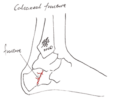
The calcaneus is the bone forming the heel. It is an important bone that provides
- a stable balancing point for the foot
- lots of area for muscle attachments (particularly the achilles tendon)
Calcaneal fractures are very common, especially in young men. They are usually due to accidents, like falls onto the feet or car accidents when the injured foot is pressed against the pedal.

Fractures may occur through the middle of the calcaneus or may involve bony edges breaking off.
Calcaneal fractures are generally classified into two categories:
- Extra-articular. These fractures do not involve any of the joints.
- Intra-articular. These fractures affect one or more joints. They often require more specialised surgery than extra-articular fractures, and are associated with more pain and dysfunction later on. The picture above shows an intra-articular fracture as the break extends up to the joint with the bone above (Talo-Calcaneal joint).
The calcaneus is also vulnerable to stress fractures, or subtle breaks in the weak bone.Like many fractures in the foot, there is a long period of not being able to stand on the foot during recovery, from 4 weeks up to 3 months. This can be frustrating, but long-term ankle function depends on the alignment of calcaneus with both the leg and the foot. Allowing the bones time to heal in place is consequently essential to recovery.
Symptoms
When the calcaneus is broken suddenly, as in car accidents or falls, the symptoms include:
- Pain around the heel and ankle
- Swelling, deformity and irritation of the skin over the heel
- Bruising
- Inability to put weight on the foot
- Tingling or numbness in the foot.
When stress fractures develop, the symptoms tend to be more subtle, gradually getting worse over time. This may make running, then walking difficult but swelling, bruising and deformity will usually be minimal.
Causes
As with many types of fractures, the causes and risk factors can be divided into two types:
- Large forces on strong bones
- Calcaneal fractures are common among sports people who sustain high-impact falls and trauma.
- Other common causes of high-energy fractures are car accidents and falls onto the feet.
- Small forces on weak bones
- Osteoporosis and advanced age
- Medications and other drugs; most commonly, people who need to take high doses of steroids for long periods of time are at risk of developing weak bones.
Risk Factors
You have a higher risk of sustaining a calcaneal fracture when you have weak bones (osteoporosis).
Performing extreme sports or high-impact sports also increase the risk of a heel fracture.
Investigations
Calcaneus fractures can often be diagnosed by a doctor or surgeon on examination, but will require a series of ankle X-Rays to confirm the diagnosis. Your radiographer may ask you to stand in a number of different positions while they take the X-Rays. This is important as there are a number of different joints associated with the calcaneus, and the integrity of each needs to be checked.
CT scans are often used when an operation is considered necessary. This scan gives more information about the exact pattern of the fractures and provides some detail about the soft tissues around the bone.
Complications
Common complications of calcaneus fractures include:
- Post-traumatic Arthritis. Any fracture near a joint will increase the risk of arthritis developing in that joint in later life. This is especially true of breaks that involve the joints around the calcaneus ( intra-articular fractures). If the arthritis is severe, you may need further surgery.
- Infection. Any surgery carries a risk of infection. For more information, see Complications of Surgery.
- Failure of the heel pad to heal. The calcaneus is protected from contact with the ground by a pad of specialised fat. This tough tissue is able to cushion the calcaneus as you put your weight through your foot while walking. Unfortunately, the fat pad does not heal well after being injured. This can lead to ongoing pain and difficulty walking.
- Nerve Injury or Entrapment. Nerve damage can be due to the accident that caused the fracture, the swelling after the fracture or a complication of surgery. Injury surrounding nerves can cause pins and needles or numbness over one side of the foot. This is usually temporary.
- Compartment syndrome. This occurs when swelling around the injured joint becomes so severe that blood vessels are squashed. The low blood supply causes injury to the nerves and other soft tissues. If you have this complication, your surgeon may need to perform an operation called a ‘fasciotomy’ to release the pressure.
- Injury to the Achilles tendon.
- Irritation from the cast.
Treatment
Initial Treatment for Calcaneal Fractures
Many fractures of the heel bone are complicated by significant swelling. To bring the swelling down and facillitate healing of the tissues, first aid treatment of the fracture includes:
- Protect your injured foot from further injury.
- Rest: Stay off the injured foot. Walking may cause further injury.
- Ice: Apply an ice pack to the injured area, placing a thin towel between the ice and the skin. Use ice for 20 minutes and then wait at least 40 minutes before icing again.
- Compression: An elastic wrap can be used to control swelling (but only allow a professional to apply a compression bandage, as inappropriate compression can impair blood supply).
- Elevation: The foot should be raised slightly above the level of your heart to reduce swelling.
These simple measures are often very effective.
Treatment in the Hospital
Treatments for calcaneus fractures depends on the type and severity of the fracture. If the fracture is not severe, a cast can be used to immobilise the foot and allow the bones to heal. More often, surgery is required.
Types of surgery for calcaneal fractures include:
- Plates and screws to fix the bones in place
- Arthrodesis (fusion). This type of surgery is dramatic and only recommended for severe fractures, or when other types of surgery have failed. Arthrodesis is very good at stabilising joints, but reduces the range of movement. If the joints around the calcaneus are fused in place, the normal stressors placed on the bones during walking changes. This can lead to arthritis and difficulty walking later in life.
For more information on treatment see Surgery for Calcaneal Fractures.
How long will the fracture take to heal?
If everything goes well and the fracture is relatively uncomplicated, the heel bone should take eight to twelve weeks to become solid bone. Over the following months the bone will continue to strengthen into near-normal bone.
However, complicated fractures, especially in those cases where the bone has shattered rather than just broken will take longer. Furthermore, fractures that become infected will need further treatment and may take much longer.
What about rehabilitation and physiotherapy?
It is important to remember that the bone may heal well, but the other structures around the ankle, including the tendons, muscles and especially the fatty heal pad may take months to heal. This includes the less severe fractures that do not require surgery – wearing a cast for a number of weeks will cause the muscles and tendons to become weak and stiff.
Rehabilitation can consequently cover weeks to months after the surgery or, if no surgery was performed, removal of the cast. Your physiotherapist will be able to discuss the need to keep the muscles around your joint strong and flexible.
Seeking Advice
Your Family Doctor (GP)
Your Family Doctor will be able to diagnose and help treat your problem. He or she will be able to
- tell you about your problem
- advise you of the best treatment methods
- prescribe you medications
- and if necessary, refer you to Specialists (Consultants) for further treatment
You may initially seek treatment for a broken heel in an emergency room or urgent-care clinic. If the pieces of broken bone aren’t lined up properly to allow healing with immobilisation, you may be referred to a doctor specialising in orthopaedic surgery.
What you can do
You may want to write a list that includes:
- Detailed descriptions of your symptoms
- Information about medical problems you’ve had
- Information about the medical problems of your parents or siblings
- All the medications and dietary supplements you take
- Questions you want to ask the doctor
Prevention
Preventing weakness of the bones is very important. If you are at risk of developing osteoporosis (for example, if you are female and over 55 yrs old) you should talk to your GP.
Preventing Complications of Calcaneal Fractures
- Elevation both immediately after the injury and after surgery will help keep swelling down and minimise the development of short-term complications. .
- Arthritis can develop in any of the joints involved in walking (hips, knees, feet and lower back) if you develop an abnormal walking pattern after the fracture. If your surgeon thinks that this is likely in your case, he may refer you on to a orthotist or podiatrist who can discuss if you need shoe inserts or orthotics.
F.A.Q. | Frequently Asked Questions
How long before I can start putting weight on my foot?
This depends on both the type of fracture and the treatment involved. You will therefore need to discuss this with your surgeon both before and after the surgery.
If surgery is NOT performed and you are put in a cast, you will generally need to be non-weight-bearing for up to 3 months. When surgery is performed, you will need to remain non-weight-bearing for the first 4 weeks at least. After this, your surgeon will advise when you can start to put some (not all) weight through your foot (see partial non-weight-bearing).
Will I have good function in my foot?
This depends on the type of fracture and how it was treated, and varies so much between individuals that you will need to discuss this with your surgeon. Often, those who are treated with surgery have better long-term function than those treated with just a plaster cast, however if you have only a minor break then a simple plaster cast will leave you with excellent function once the bone has healed.
When can I drive again?
This will vary from case to case, and will depend on you, your injury and your insurance. In general, driving is seen as a ‘weight-bearing-activity’, meaning that when you are starting to take more weight through your foot you are getting closer to being able to drive.
A few things to keep in mind:
- You can’t drive unless you can perform an emergency stop. In some cases, you may need to demonstrate this in order to be insured on the road.
- Many people feel well enough to drive long before they are considered officially safe to drive. While it may be tempting to drive during this time, it’s important to remember that if you have a crash, your insurance is likely to be invalid, and you may be liable to damages to both your own and the other party’s car.
More Information
More information on fractures, relevant treatments and support services can be found at the following sites:
- Patient information, forums and stories
- Real Time Health – videos and stories from patients
- Virtual Medicine Centre
- MyDr Australia
- Physiotherapy and Rehabilitation
- Physiotherapy Choices – a site offering explanations of the findings in the latest research available
- Centre for Evidence Based Physiotherapy
- Allied Health Evidence
- Healthy Living & Other Useful Sources
- Health Insite – an Australian government website providing links to information on the net
- Preventing Sports Injuries on Health Insite
- Better Health Channel
- Department of Health and Ageing
- Your Health
- Medicines.org.au
- Consumers Health Forum
- Health Insite – an Australian government website providing links to information on the net
Medical sources on Fifth Metatarsal Fractures:
The latest research on fifth metatarsal fractures is accessible through the Cochrane Library and PubMed Database.
References
Matherne, T. H., et al., ‘Calcaneal Fractures: What the Surgeon Needs to Know’, Current Problems in Diagnostic Radiology, Vol 36, Iss 1, Jan-Feb 2007, pp 1-10.
McCormack, A. D., ‘Chapter 32: Calcaneal Fractures’, Treatment and Rehabilitation of Fractures, Hoppenfield S., Murthy, V., (Eds), Lippincott Williams & Wilkins, Philadelphia, USA, 2000.
Parvizi, J., Kim, G. K., ‘Calcaneal Fractures’, High Yield Orthopaedics, 2010, pp 72-73.
